In radiation therapy (RT), knowledge of precise boundary locations of objects and the target tumor is essential for devising an irradiation plan for delivering the required dose to the tumor and minimizing radiation dose to normal and critical anatomical structures. The first step in RT planning is therefore to define object and tumor contours on planning CT, MRI, or PET/CT images. This process of delineating contours is still performed with low levels of automation due to lack of highly automated commercial contouring software. This deteriorates therapy planning due to several reasons. (1) Manual contouring is error-prone, subject to intra- and inter-observer variations, labor-intensive, and results in suboptimal throughput. (2) In advanced and modern conformal radiation techniques like intensity modulated radiotherapy (IMRT) and proton beam radiation therapy (PBRT), anatomic changes taking place during a 5-8 week course of treatment are not accounted for due to manual labor involved in contouring. (3) Such changes can significantly affect the total dose delivered to the tumor and normal surrounding organs and are particularly important to account for when treating malignancies that are highly radiosensitive. The ability to automatically delineate changes in patient anatomy during therapy could establish adaptive radiotherapy as a routine clinical treatment protocol for reducing the total dose to normal critical structures and delivering highly precise radiotherapy to tumors, which can lead to improved patient outcomes and cost-saving.
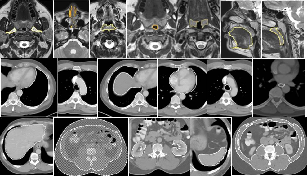
Based on the AAR technology, we are developing software tools for contouring anatomic objects at a high level of automation on diagnostic CT, low-dose CT, and MR images. An example illustrating automated AAR contouring of numerous anatomic objects on diagnostic CT in the thorax and abdomen and on MR images in the neck is presented below.
References:
1. Udupa JK, Odhner D, Zhao L, Tong Y, Matsumoto MM, Ciesielski KC, Falcao AX, Vaideeswaran P, Ciesielski V, Saboury B, Mohammadianrasanani S, Sin S, Arens R, Torigian DA. Body-wide hierarchical fuzzy modeling, recognition, and delineation of anatomy in medical images. Med Image Anal. 2014;18(5):752-71. doi: 10.1016/j.media.2014.04.003. PubMed PMID: 24835182; PMCID: PMC4086870.
2. Wu X, Udupa JK, Tong Y, Odhner D, Pednekar GV, Simone CB, 2nd, McLaughlin D, Apinorasethkul C, Apinorasethkul O, Lukens J, Mihailidis D, Shammo G, James P, Tiwari A, Wojtowicz L, Camaratta J, Torigian DA. AAR-RT - A system for auto-contouring organs at risk on CT images for radiation therapy planning: Principles, design, and large-scale evaluation on head-and-neck and thoracic cancer cases. Med Image Anal. 2019;54:45-62. doi: 10.1016/j.media.2019.01.008. PubMed PMID: 30831357; PMCID: PMC6499546.