Systematic assessment of regional lymph nodes in cancer patients in a standardized manner is important for disease staging and for prognostication of patient outcome. For certain body regions like the thorax, standards such as the one proposed by the International Association for the Study of Lung Cancer (IASLC) for defining nodal stations are available. While this formulation is helpful in standardizing a means of interpreting and reporting thoracic lymph node disease sites, it still leaves the radiologist with the arduous task of following the detailed specifications and finding the nodal stations and zones on images subjectively. Our hypothesis is that if the lymph node stations can be identified automatically during image interpretation, then the acceptance of the standard and the consistency of its interpretation will be greatly facilitated, which may rapidly promote standardized reporting. In this project, we not only develop nodal station definitions for body regions where definitions do not exist currently, but also employ the AAR technology for automatically localizing the lymph node stations in different body regions in CT, PET/CT, and MR images and subsequently quantify lymph node disease burden.
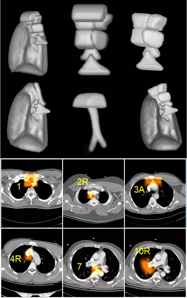
An example from such an implementation for identifying the IASLC defined thoracic nodal stations is shown below where 3D renditions of the fuzzy models of some stations together with some anatomic objects are displayed as well as the AAR-localized zones overlaid on CT slice images.
AAR-LN-DQ: Automatic anatomy recognition based disease quantification in thoracic lymph node zones via FDG PET/CT images without Nodal Delineation
Purpose: The derivation of quantitative information from medical images in a practical manner is essential for quantitative radiology (QR) to become a clinical reality, but still faces a major hurdle because of image segmentation challenges. With the goal of performing disease quantification in lymph node (LN) stations without explicit nodal delineation, this paper presents a novel approach for disease quantification (DQ) by automatic recognition of LN zones and detection of malignant lymph nodes within thoracic LN zones via positron emission tomography/computed tomography (PET/CT) images. Named AAR-LN-DQ, this approach decouples DQ methods from explicit nodal segmentation via an LN recognition strategy involving a novel globular filter and a deep neural network called SegNet. Method: The methodology consists of four main steps: (a) Building lymph node zone models by automatic anatomy recognition (AAR) method. It incorporates novel aspects of model building that relate to finding an optimal hierarchy for organs and lymph node zones in the thorax. (b) Recognizing lymph node zones by the built lymph node models. (c) Detecting pathologic LNs in the recognized zones by using a novel globular filter (g-filter) and a multi-level support vector machine (SVM) classifier. Here, we make use of the general globular shape of LNs to first localize them and then use a multi-level SVM classifier to identify pathologic LNs from among the LNs localized by the g-filter. Alternatively, we designed a deep neural network called SegNet which is trained to directly recognize pathologic nodes within AAR localized LN zones. (d) Disease quantification based on identified pathologic LNs within localized zones. A fuzzy disease map is devised to express the degree of disease burden at each voxel within the identified LNs to simultaneously handle several uncertain phenomena such as PET partial volume effects, uncertainty in localization of LNs, and gradation of disease content at the voxel level. We focused on the task of disease quantification in patients with lymphoma based on PET/CT acquisitions and devised a method of evaluation. Model building was carried out using 42 near-normal patient datasets via contrast-enhanced CT examinations of the thorax. PET/CT datasets from an additional 63 lymphoma patients were utilized for evaluating the AAR-LN-DQ methodology. We assess the accuracy of the three main processes involved in AAR-LN-DQ via fivefold cross validation: lymph node zone recognition, abnormal lymph node localization, and disease quantification. Results: The recognition and scale error for LN zones were 12.28 mm +- 1.99 and 0.94 +- 0.02, respectively, on normal CT datasets. On abnormal PET/CT datasets, the sensitivity and specificity of pathologic LN recognition were 84.1% +-0.115 and 98.5% +-0.003, respectively, for the g-filter SVM strategy, and 91.3% +- 0.110 and 96.1% +- 0.016, respectively, for the SegNet method. Finally, the mean absolute percent errors for disease quantification of the recognized abnormal LNs were 8% +- 0.09 and 14% +- 0.10 for the g-filter-SVM method and the best SegNet strategy, respectively. Conclusions: Accurate disease quantification on PET/CT images without performing explicit delineation of lymph nodes is feasible following lymph node zone and pathologic LN localization. It is very useful to perform LN zone recognition by AAR as this step can cover most (95.8%) of the abnormal LNs and drastically reduce the regions to search for abnormal LNs. This also improves the specificity of deep networks such as SegNet significantly. It is possible to utilize general shape information about LNs such as their globular nature via g-filter and to arrive at high recognition rates for abnormal LNs in conjunction with a traditional classifier such as SVM. Finally, the disease map concept is effective for estimating disease burden, irrespective of how the LNs are identified, to handle various uncertainties without having to address them explicitly one by one.
References:
1. Xu G, Udupa JK, Tong Y, Odhner D, Cao H, Torigian DA. AAR-LN-DQ: Automatic anatomy recognition based disease quantification in thoracic lymph node zones via FDG PET/CT images without Nodal Delineation. Med Phys. 2020;47(8):3467-84. doi: 10.1002/mp.14240. PubMed PMID: 32418221.